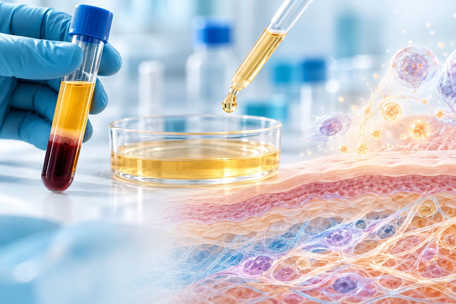
How can the biological potential of Platelet-Rich Plasma (PRP) be evaluated using human skin models?
At BioToSkin, we conducted a comprehensive ex vivo study to investigate the effects of a topical formulation combined with PRP on human skin.
By integrating skin hydration measurements, collagen and elastin immunostaining, and growth factor quantification, the study demonstrated the value of a multi-endpoint experimental approach for evaluating innovative skin regeneration technologies.
The results revealed significant improvements in superficial skin hydration, encouraging changes in structural skin markers, and measurable growth factor release throughout the experimental period.
PRP: a promising approach in skin regeneration research
Platelet-Rich Plasma (PRP) is increasingly investigated in aesthetic medicine and regenerative applications because of its concentration of platelets and associated growth factors.
These factors participate in biological processes related to tissue repair, extracellular matrix remodeling and cellular renewal.
Among the most relevant molecules are:
- PDGF-BB (Platelet-Derived Growth Factor-BB): associated with tissue repair and extracellular matrix remodeling.
- EGF (Epidermal Growth Factor): involved in epidermal renewal and cellular proliferation.
Evaluating PRP-based technologies requires more than investigating a single biomarker. A comprehensive approach should examine both growth factor availability and biological responses in skin tissue.
This was the strategy adopted by BioToSkin.
Study design: combining functional, structural and biochemical endpoints
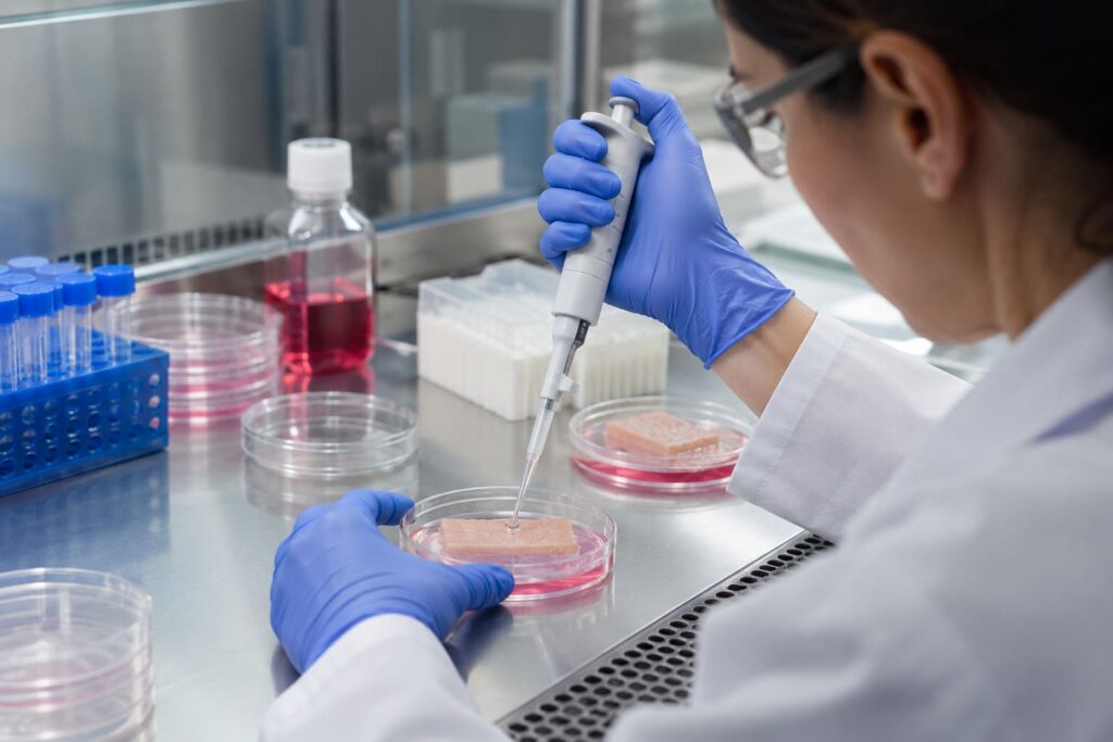
The investigation was conducted using human skin explants maintained in ex vivo culture.
Three experimental groups were evaluated, with three explants per group:
- Untreated skin
- Skin treated with a topical formulation
- Skin treated with the topical formulation combined with PRP
The study combined several complementary techniques.
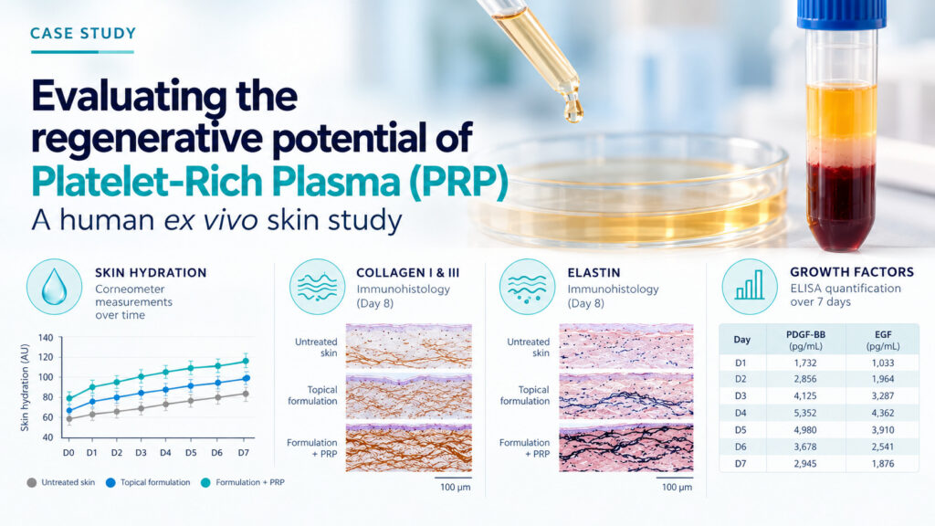
Skin hydration — Corneometer
Superficial hydration was monitored daily over seven days to assess functional changes in the skin.
Collagen I and III — Immunohistology
Skin samples were analyzed at days 2, 6 and 8 to investigate changes in structural proteins associated with dermal organization and firmness.
Elastin — Immunohistology
Elastin staining was assessed to investigate the response of a major structural component associated with skin elasticity.
Growth factors — ELISA
PDGF-BB and EGF were quantified over seven days following stimulation of PRP with the topical formulation.
This experimental design allowed BioToSkin to evaluate several dimensions of skin response within a coordinated study.
Result 1: significant improvement in skin hydration
One of the most notable findings was the improvement in superficial skin hydration observed with the formulation–PRP combination.
After seven days, the mean hydration measurements were:
| Experimental group | Hydration at day 7 |
|---|---|
| Untreated skin | 80.00 AU |
| Topical formulation alone | 93.33 AU |
| Topical formulation + PRP | 106.67 AU |
Compared with the untreated group, the formulation–PRP combination showed approximately 33% higher superficial hydration.
It also produced a value approximately 14% higher than the formulation alone.
The dedicated day-7 statistical analysis identified significant differences:
- Formulation + PRP vs. untreated skin: p < 0.001
- Formulation + PRP vs. formulation alone: p = 0.006
These findings provided quantitative evidence of an enhanced hydration response under the experimental conditions.
Importantly, the study demonstrated that the human skin explant model could detect and quantify treatment-related differences over time.
Result 2: increased collagen I and III staining
Beyond hydration, BioToSkin investigated the response of major structural proteins involved in maintaining skin integrity.
Collagen I
Immunohistological observations showed increased collagen I staining in skin explants treated with the formulation–PRP combination.
A slight increase was observed at day 2, followed by more pronounced differences at days 6 and 8.
The staining changes were observed across several skin compartments, including the epidermis, superficial dermis and deep dermis.
Collagen III
Increased collagen III staining was also observed at day 2, particularly in the epidermis and superficial dermis.
These results demonstrated the ability of the experimental model to reveal treatment-associated changes in structural skin markers.
The findings were qualitative and based on immunohistological observations rather than direct quantification of newly synthesized collagen.
Result 3: enhanced elastin staining
Elastin is an essential component of the extracellular matrix, contributing to the skin’s ability to recover its shape following deformation.
The study revealed more pronounced elastin staining in skin explants treated with the formulation–PRP combination compared with untreated skin and the formulation alone.
Changes were visible from day 2, particularly in the superficial and deep dermis.
At day 8, the difference in elastin staining was especially marked across the examined skin compartments.
These observations complemented the collagen findings and provided further evidence of measurable structural responses in the ex vivo skin model.
By combining several immunohistological markers, BioToSkin was able to characterize changes in skin structure across different tissue compartments and experimental time points.
Result 4: measurable growth factor release over seven days
Another important achievement of the study was the successful monitoring of PRP-derived growth factors.
Using ELISA assays, BioToSkin quantified PDGF-BB and EGF following stimulation of the PRP preparation.
The measured concentrations ranged from:
| Growth factor | Measured concentration range |
|---|---|
| PDGF-BB | 1,732–5,352 pg/mL |
| EGF | 1,033–4,362 pg/mL |
Both growth factors remained measurable throughout the seven-day monitoring period.
These results demonstrated that the experimental approach could characterize growth factor release over time.
The ability to combine biochemical measurements with tissue-based analyses represents an important advantage when evaluating PRP-related technologies.
Why this study highlights BioToSkin’s expertise
The main achievement of this investigation was not limited to one positive endpoint.
It was the successful integration of multiple complementary techniques to characterize the biological response of human skin to an innovative treatment approach.
The study generated four important categories of evidence:
- Functional evidence: quantitative differences in superficial skin hydration, including statistically significant results.
- Structural evidence: increased staining of collagen I, collagen III and elastin in treated skin explants.
- Biochemical evidence: measurable release of PDGF-BB and EGF over seven days.
- Temporal evidence: characterization of skin responses and growth factor availability at several experimental time points.
This combination provided a more comprehensive assessment than any single measurement could have delivered.
It also illustrated the flexibility of human ex vivo skin models for investigating emerging technologies in cosmetics, dermocosmetics and aesthetic medicine.
Translating scientific questions into measurable endpoints
Innovative skin treatments frequently involve complex biological mechanisms.
Evaluating these technologies requires selecting relevant biomarkers, establishing appropriate controls and using complementary analytical techniques.
At BioToSkin, our approach is to design experimental protocols around the scientific question being investigated.
Depending on the objective, studies can combine:
- Human skin explant culture
- Functional skin measurements
- Histology and immunohistochemistry
- ELISA-based biomarker quantification
- Skin penetration and distribution studies
- Customized experimental kinetics
This integrated strategy enables the investigation of skin responses at functional, structural and molecular levels.
Conclusion: a successful multi-endpoint evaluation of PRP on human skin
This study demonstrated the feasibility and scientific value of evaluating a PRP-based topical approach using human ex vivo skin explants.
The investigation generated several complementary findings:
- Significant hydration differences, with the formulation–PRP group reaching 106.67 AU at day 7.
- Increased collagen I, collagen III and elastin staining, providing encouraging evidence of structural skin responses.
- Measurable growth factor release, with PDGF-BB and EGF detected throughout seven days of monitoring.
Together, these results demonstrate the ability of BioToSkin’s experimental platform to investigate complex skin regeneration technologies using complementary biological endpoints.
Although exploratory and based on a limited number of explants, the study provides a strong methodological example of how human skin models can generate meaningful preclinical evidence.
At BioToSkin, we transform complex scientific questions into measurable biological evidence.
Are you developing an innovative cosmetic, dermocosmetic or aesthetic treatment?
Contact BioToSkin to explore how our customized human skin models can support your research and product development.