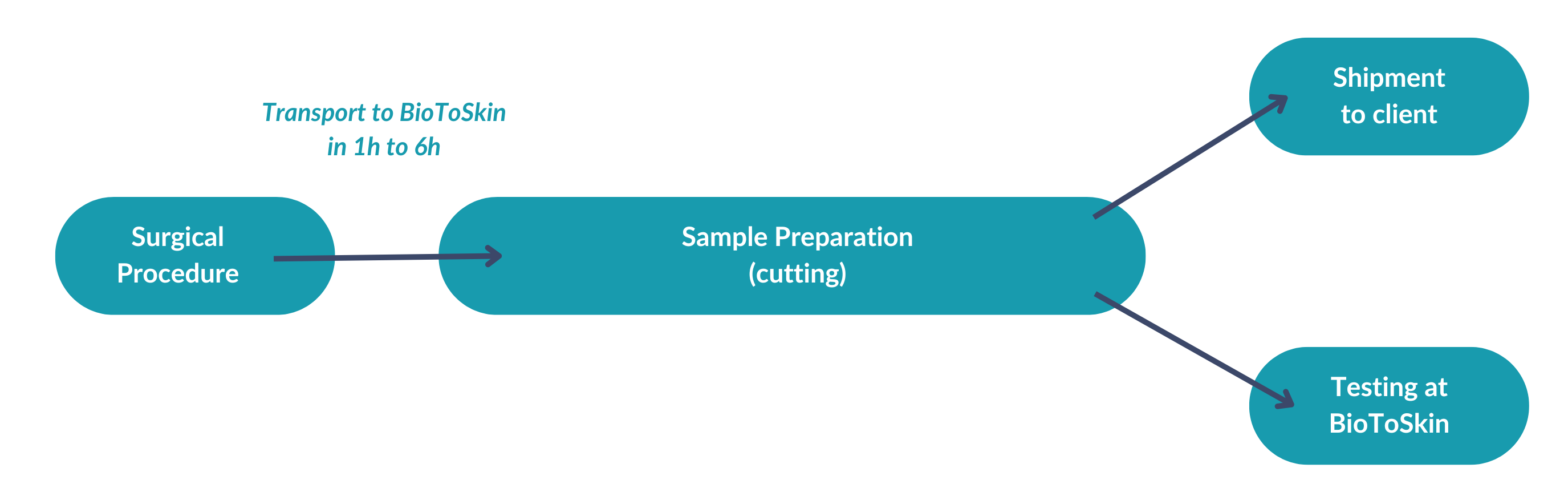
Ex vivo methods of skin penetration study by Franz cellprovide pharmacokinetic information on the diffusion of a substance through the skin. These methods are particularly suitable for comparing the transport of chemicals in and through the skin, for different preparations, but they also provide useful models for the assessment of percutaneous absorption in humans.
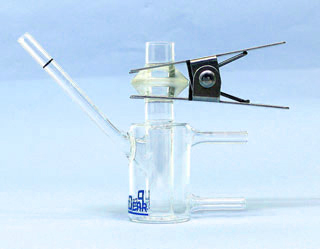
BioToSkin has several automated or manual diffusion systems (Microette 18 cells Hanson, Permagear…) per Franz cell in order to offer a solution adapted to customers. We also have the option of working on synthetic (Strat M, Omnipore, etc.) or biological membranes.
In order to guarantee a higher quality of the assay, we carry out the HPLC analyzes within our structure with proven chromatography methods.
Click on the image to enlarge
Microdialysis
Ex vivo microdialysis is a method of exploration on human explant taking into account the cutaneous metabolism. First used in neuropharmacological research, its application in cutaneous biology and pharmacology is more recent but in full expansion.
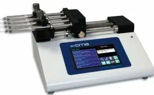
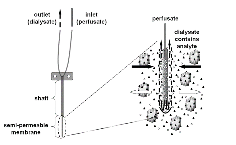
The basic principle of this system is shown schematically in the image opposite: a semi-permeable fiber (permeable to water and small compounds), introduced into a fabric, will recover the molecule sought according to a concentration gradient. This fiber is connected to a micro-pump, which allows infusion at a defined flow rate of a physiological liquid. This method makes it possible to monitor the production of both endogenous and exogenous molecules (pharmacokinetic study).
This exploration method is the closest to in vivo conditions. For example, it is possible to monitor the production of molecules responsible for oxidative stress or inflammation.
Exposome and Environmental Stress:
Our ex vivo human skin models allow the investigation of different environmental stressors affecting the skin, such as pollution, UV exposure, blue light, oxidative stress and inflammatory mediators.
These approaches make it possible to assess the ability of an active ingredient, raw material or finished product to limit skin alterations induced by the cutaneous exposome. Biological responses can be analysed through different markers, including inflammation, oxidative stress, tissue viability, morphological alterations, matrix degradation and activation of cellular defence pathways.
These models are particularly relevant for claims related to protection, prevention of environmental skin ageing, anti-pollution efficacy and reinforcement of skin resilience.
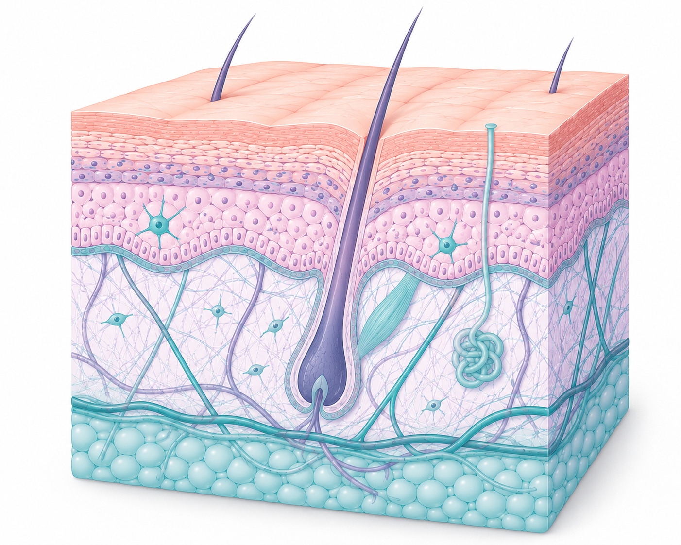
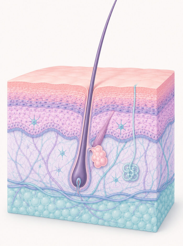
Skin Barrier and Repair
Ex vivo human skin is a relevant model to study skin barrier integrity and its capacity to recover after controlled alteration.
We can evaluate the effect of active ingredients or formulations on different parameters related to barrier function, including tissue organisation, epidermal marker expression, cellular cohesion and skin differentiation markers such as filaggrin, claudin-1, loricrin or involucrin.
These studies help document the repairing, regenerative or protective properties of a product, particularly for claims related to skin repair, barrier reinforcement or recovery after stress.
Anti-pollution effect
Objectification studies of anti-pollution claims make it possible to quantify the protective effect of the formulation against identified pollutants on skin explants kept alive.
We have developed models using the most widespread pollutants (PM2.5, PM10, PAHs, etc.) in order to analyze markers of oxidative stress (lipid peroxidation, protein carboxylation, immunohistology, AhR, etc.)
Hair Fibre & Scalp Ex Vivo Studies
BioToSkin also offers ex vivo approaches dedicated to hair and scalp-related applications.
Using human hair fibres and, when available, hair-bearing skin explants, we can assess the effects of ingredients or finished products on fibre integrity, surface morphology, damage repair, protection against environmental or heat-induced stress, and scalp–hair interface biology.
These models are particularly relevant for claims related to hair repair, protection, conditioning, anti-pollution efficacy and post-treatment recovery