

Injectable fillers and biostimulators are no longer assessed solely for their volumizing effect. Their interaction with the dermal microenvironment, early inflammation and extracellular matrix remodeling are becoming key characterization parameters.
Microdialysis adds an essential dimension: the local kinetics of soluble molecules within the extracellular compartment.
1. Why Filler Biology Requires a Dynamic Approach
Injectable products used in aesthetic medicine include materials with very different properties: hyaluronic acid (HA), poly-L-lactic acid (PLLA), poly-D,L-lactic acid (PDLLA), calcium hydroxyapatite (CaHA), polycaprolactone and hybrid formulations.
Their performance does not depend solely on their rheological properties or their ability to restore volume. The literature also describes interactions with fibroblasts, macrophages, TGF-β pathways and extracellular matrix synthesis [4–7].
For R&D teams, this raises a simple question that is difficult to address with a single endpoint: How does the tissue respond around the injected product, and how does this response evolve over time?
2. The Principle of Cutaneous Microdialysis
Microdialysis relies on a probe equipped with a semi-permeable membrane, positioned within the tissue and perfused at a low flow rate.
Soluble molecules present in the interstitial fluid diffuse across the membrane and are collected in the dialysate. This technique therefore provides access to the local extracellular compartment and can be used in both clinical and preclinical settings, including ex vivo models [1,2].
The key advantage is the ability to collect several successive fractions from the same tissue.
Instead of obtaining only a terminal measurement, microdialysis provides a concentration–time curve, making it possible to identify an early response, a peak, a decline phase or a prolonged response.
3. What Microdialysis Adds Compared with Conventional Methods
Microdialysis does not replace histology, immunohistochemistry or molecular analyses. It addresses a different question. A culture supernatant provides information on what is released by the entire explant. A tissue homogenate provides information on the overall content of a sample. IHC or IF localizes a marker within the tissue. Microdialysis, by contrast, repeatedly samples the extracellular environment within a defined area.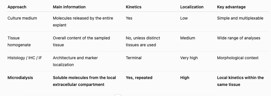
4. Why This Approach Is Particularly Relevant for Fillers and Biostimulators
Following injection, the product creates a local interface between the biomaterial and the tissue.
This area concentrates interactions between the extracellular matrix, immune cells, fibroblasts and soluble mediators. However, a highly localized response may become diluted when analyzing the culture medium of an entire explant.
For a filler, microdialysis can therefore be used to characterize:
- the early inflammatory response around the injectable deposit;
- the kinetics of mediators associated with biostimulation or remodeling;
differences in profiles between formulations, concentrations or crosslinking technologies; - the effect of product combinations or combined protocols;
- the relationship between an early biological response and tissue endpoints measured at a later stage.
This approach is particularly relevant for materials claiming biostimulatory activity.
Studies on PLLA have, for example, demonstrated activation of the TGF-β/Smad pathway associated with increased collagen synthesis by fibroblasts [5]. Other studies have also shown that different HA fillers can induce distinct fibroblast responses, particularly regarding collagen, elastin and TGF-β1 [6].
5. Which Biomarkers Should Be Monitored?
Biomarker selection should be guided by the expected mechanism of action and by the actual recovery capacity of the probe.
For injectable products, several families of markers may be considered.
5. Quels biomarqueurs suivre ?
Le choix des biomarqueurs doit être guidé par le mécanisme d’action attendu et par la capacité réelle de récupération de la sonde. Pour les produits injectables, plusieurs familles de marqueurs peuvent être envisagées.
The cutaneous microdialysis literature notably documents the monitoring of cytokines such as IL-1β, IL-6 and IL-8, with kinetic profiles that may include several phases [3].
6. A Platform Already Applied at BioToSkin
At BioToSkin, microdialysis has been adapted to human skin explants to investigate the local response to injectable products.
In a recent internal series, several filler conditions were compared over 24 hours, monitoring TGF-β, IL-6, IL-8 and β-galactosidase, with calculation of the area under the curve (AUC 0–24 h).
These unpublished internal data demonstrate the practical value of the platform for comparing biological profiles across formulations without being restricted to a single measurement time point.
The objective is not to summarize a product using a single marker, but rather to build a profile encompassing: response intensity, timing, duration, and the balance between inflammation and biostimulation.
7. The Value of Combining Microdialysis and Histology
The power of the approach increases when microdialysis is combined with terminal analyses.
Microdialysis provides information on mediators released during the experiment, while histology and IHC/IF allow assessment of tissue architecture and structural responses.
- During the study: dialysate fractions for IL-6, IL-8, TGF-β or other biomarkers.
- At the end of the study: histology, collagen I/III, elastin, α-SMA, macrophages, MMP/TIMP or other targeted endpoints.
- Integrated analysis: linking an early signal to subsequent tissue remodeling.
8. Limitations to Consider
Analyte recovery depends on several parameters, including the molecular weight cut-off of the membrane, perfusion flow rate, collection duration, probe depth, membrane length and the potential adsorption of certain molecules [2].
Large proteins may exhibit lower recovery than smaller molecules.
Probe implantation also constitutes a local perturbation. An equilibration period, appropriate controls, and strict standardization of probe position and injection site are therefore essential.
Microdialysis should be regarded as a specialized measurement tool integrated into a coherent experimental design.
9. Toward a More Translational Characterization of Injectable Products
2D cell models are valuable for dissecting mechanisms. Clinical studies remain essential for demonstrating performance in humans.
Between the two, ex vivo human skin preserves tissue architecture and interactions between multiple cellular compartments while allowing experimental conditions to be tightly controlled.
The combination of: ex vivo human skin + intradermal injection + microdialysis + tissue analyses therefore represents a particularly relevant strategy for comparing fillers, skin boosters and biostimulators, documenting their local biological response and generating mechanistic data complementary to in vitro and clinical studies.
Conclusion
For developers of injectable products, the question is no longer simply:
“Does the product increase a marker at 24 hours?”
but rather:
“What biological response does it trigger around the injection site, with what intensity and according to what kinetics?”
Ex vivo cutaneous microdialysis addresses this question by directly sampling the local extracellular compartment.
Combined with histology, IHC/IF and biomarker assays, it provides a more dynamic and mechanistic understanding of the tissue response to fillers and biostimulators
Are you developing a filler, skin booster or biostimulator?
BioToSkin can develop an ex vivo human skin model combining injection, microdialysis, biomarkers, histology and immunohistochemistry, tailored to the product’s mechanism of action and the level of evidence required.Contact-us.Scientific References
1. Baumann KY, Church MK, Clough GF, et al. Skin microdialysis: methods, applications and future opportunities—an EAACI position paper. Clin Transl Allergy. 2019;9:24. doi:10.1186/s13601-019-0262-y. PubMed
2. Rea H, Kirby B. A Review of Cutaneous Microdialysis of Inflammatory Dermatoses. Acta Derm Venereol. 2019;99(11):945–952. doi:10.2340/00015555-3223. PubMed
3. Quist SR, Wiswedel I, Quist J, Gollnick HP. Kinetic Profile of Inflammation Markers in Human Skin In vivo Following Exposure to Ultraviolet B Indicates Synchronic Release of Cytokines and Prostanoids. Acta Derm Venereol. 2016;96(7):910–916. doi:10.2340/00015555-2406. PubMed
4. Cassuto D, Bellia G, Schiraldi C. An Overview of Soft Tissue Fillers for Cosmetic Dermatology: From Filling to Regenerative Medicine. Clin Cosmet Investig Dermatol. 2021;14:1857–1866. doi:10.2147/CCID.S276676. PubMed
5. Zhu W, Dong C. Poly-L-Lactic acid increases collagen gene expression and synthesis in cultured dermal fibroblast (Hs68) through the TGF-β/Smad pathway. J Cosmet Dermatol. 2023;22(4):1213–1219. doi:10.1111/jocd.15571. PubMed
6. Varì S, Minoretti P, Emanuele E. Human dermal fibroblast response to hyaluronic acid-based injectable dermal fillers: an in vitro study. Postepy Dermatol Alergol. 2022;39(6):1088–1092. doi:10.5114/ada.2022.114927. PubMed
7. Cabral LRB, Teixeira LN, Gimenez RP, et al. Effect of Hyaluronic Acid and Poly-L-Lactic Acid Dermal Fillers on Collagen Synthesis: An in vitro and in vivo Study. Clin Cosmet Investig Dermatol. 2020;13:701–710. doi:10.2147/CCID.S266015. PubMed
Source interne : BioToSkin, données non publiées de microdialyse ex vivo sur produits injectables (suivi TGF-β, IL-6, IL-8 et β-galactosidase, AUC 0–24 h).