
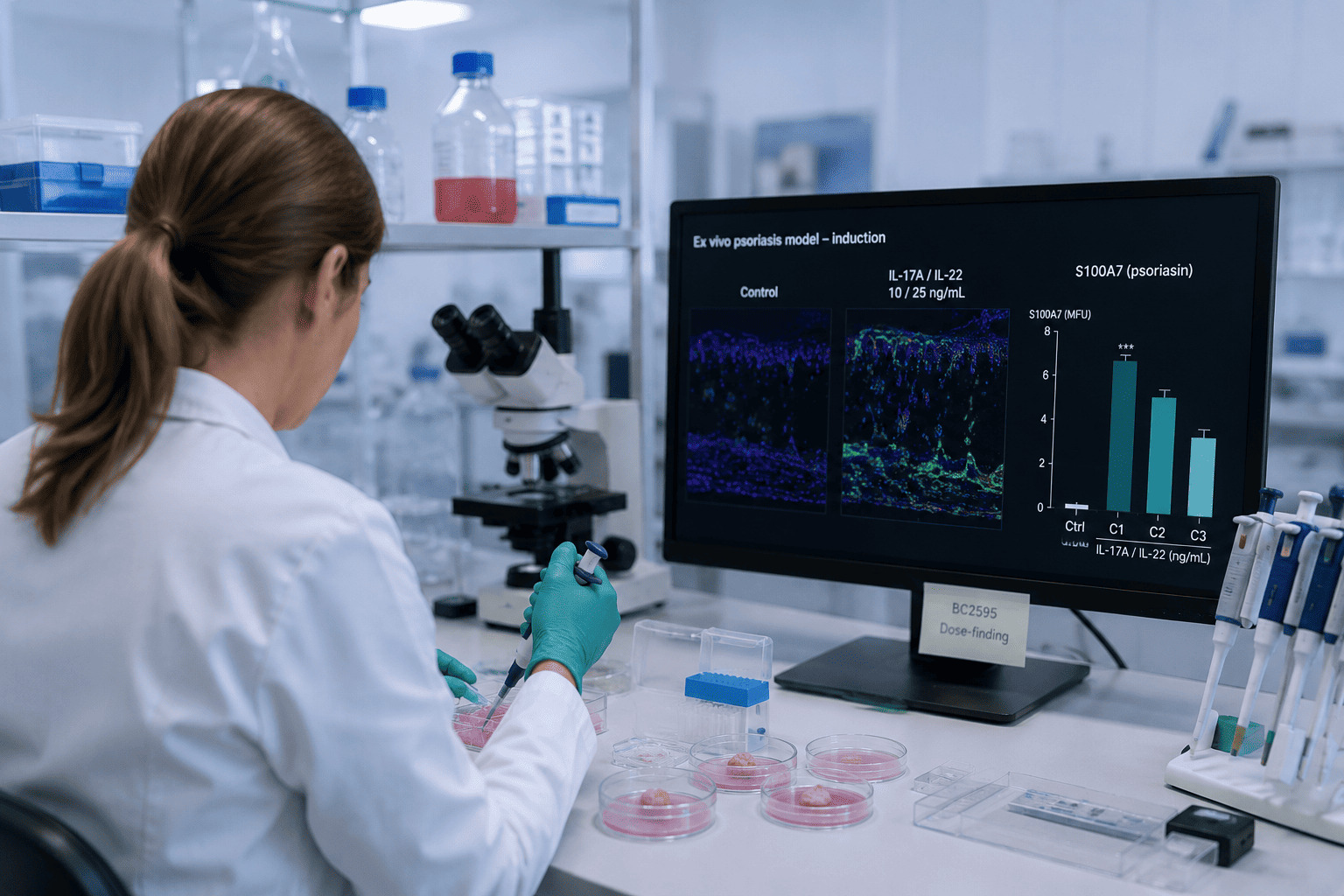
We tested three IL-17/IL-22 doses on human skin explants before running a single efficacy study.
The most aggressive cocktail produced the lowest inflammatory readout of the three. That result shaped how we build every disease model we run — and it is the reason a modest 39% induction window can still separate three topical formulations
The step most screening assays skip
The IL-23/Th17 axis is the accepted driver of psoriasis pathogenesis, and IL-17A and IL-22 are its two operative cytokines on keratinocytes (Nograles et al., 2008). Stimulating skin with an IL-17/IL-22 cocktail to obtain a psoriasis-like phenotype is, at this point, textbook.
Two things upstream of that are not. The first is the tissue: this study runs on fresh human skin from scheduled abdominoplasty surgery, collected with donor consent and put into culture within a short ischemia window — a logistics chain we operate in-house rather than working from banked or frozen material. The second is the readout: fixation, paraffin embedding, immunofluorescence and ImageJ quantification are all run under one roof, so induction, treatment and measurement stay on the same controlled pipeline. Those two capabilities are what make the dose-finding below possible in the first place.
What is not textbook is the dose. Published cocktails span a wide concentration range, and most screening assays simply adopt one and move on. We treated the dose as an experimental question in its own right: before any formulation was applied, we ran a dedicated dose-finding study on a separate donor, comparing three IL-17/IL-22 pairs against two markers — psoriasin (S100A7) and cytokeratin 16 (CK16) — with a tissue viability control (MTT) running in parallel.
The intuition going in was the obvious one: more cytokine, more inflammation, bigger window. The data said otherwise
More stimulation, less signal
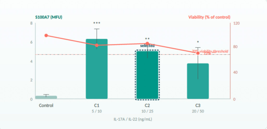
Every dose induced psoriasin significantly above a near-undetectable baseline. But the induction was strongest at the lowest dose and weakest at the highest — and the highest dose was the only one to compromise viability, at 72.1% of control (p<0.01), right at the cytotoxicity threshold.
The two curves point the same way, and the reading is hard to avoid: past a certain point you are no longer stimulating an inflammatory pathway — you are damaging the tissue that has to run it, and stressed keratinocytes make less psoriasin, not more. On a single donor the inverse dose response is a trend rather than a proven effect; what is not a trend is the viability loss at the top dose. Either way, the direction is the wrong one for anybody hoping that a bigger cytokine load buys a bigger window
We selected the intermediate pair, IL-17 10 ng/mL + IL-22 25 ng/mL. Not because it gave the biggest number — the lowest dose did — but because it was the only condition to engage both dimensions of the psoriatic phenotype (inflammatory induction and a hyperproliferative trend on CK16) while leaving the tissue intact.
An endpoint we chose because the data chose it
We measured two markers throughout. We are going to tell you about both, including the one that did not work.
S100A7 (psoriasin) performed. Near-undetectable at baseline, strongly induced, tightly reproducible.
CK16 did not. In dose-finding it rose only at the intermediate pair, and even there it fell short of significance (p = 0.13).
In the efficacy study, it did not move at all — no difference between any condition, not even control versus induction.
As a hyperproliferation marker on a 5-day explant, it is simply too slow and too variable to discriminate.
So psoriasin became the primary efficacy endpoint and CK16 stayed in the report as a complementary readout, interpreted with caution. That is a finding, not a design choice made in advance — and a screening platform that only ever tells you what worked is a platform that is not telling you everything.
Precision, not amplitude: what a 39% window can actually resolve
In the efficacy study, induction raised psoriasin from 9.92 to 13.83 a.u. — +39%, p<0.001. Read on its own, that is a modest window, and the obvious objection follows: with only 39% of headroom, can you really rank three topical creams?
You can, if your measurement is quiet enough. Here is the full readout, with the coefficient of variation for each condition:
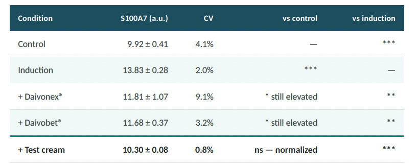
The induction window is 39%. The measurement noise across conditions runs from 0.8% to 9.1%, with a median around 3%. It is that ratio — roughly ten to one — and not the size of the window, that determines whether formulations can be separated.
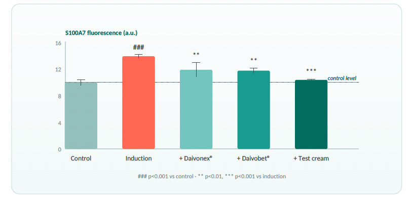
The ranking that comes out of it is specific rather than promotional. All three creams reduced psoriasin significantly.
Both reference treatments — Daivonex® (calcipotriol) and Daivobet® (calcipotriol + betamethasone) — left the marker significantly above control. The test cream alone normalized it completely (p = 0.90 vs control), outperforming Daivonex® directly (p = 0.05) and trending above Daivobet® (p = 0.07, not significant).
A viability control ran alongside every condition: no significant difference between any of them. The drop in psoriasin
is an anti-inflammatory effect, not cell loss.
What this changes for a formulation programme
The model is calibrated before your product touches it.
Dose-finding on a dedicated donor is part of the study, not an assumption inherited from a paper.
Induction stays in the physiological range.
An exaggerated model would have looked more impressive and told you less — our own dose curve points the other way
Finished formulations, applied topically at 2 mg/cm² through an intact barrier
— excipients and delivery contribute to the result, as they will in use.
Fresh human tissue and in-house analytics.
Skin sourced from scheduled surgery on a short ischemia window, with histology, immunofluorescence and image quantification run on one integrated pipeline — not outsourced, not frozen.
You get the negative results too.
CK16 did not discriminate here, and the report says so.
WHAT THIS DESIGN ESTABLISHES — AND WHAT IT DOES NOT
- Set-up and efficacy were each run on one donor, n = 3 replicates per condition. The study establishes discrimination between formulations within a donor; it does not yet quantify inter-donor variability.
- The test cream vs Daivonex® separation sits at p = 0.05 — real, but at the threshold. Confirming the ranking calls for a multi-donor design.
- CK16 was not modulated in the efficacy study. Psoriasin carries the discrimination here; hyperproliferation would need a longer or differently stressed model.
- The natural next step is 3–4 donors at the calibrated dose, which converts a strong singledonor signal into a claim that survives review.
Supporting the development of next-generation dermatological products
Human ex vivo explants are increasingly recognized as the bridge between conventional in vitro assays and clinical studies, precisely because they retain the tissue architecture that a monolayer cannot (Ubago-Rodríguez et al., 2024).
But an explant model is only as good as its calibration — and calibration is the part that rarely gets published.
Ours is on this page, including the dose that failed and the marker that did not work. If a preclinical model is going to inform a real formulation decision, that is the level at which it should be discussed.
Evaluate your topical formulation in a calibrated human psoriasis
model
Talk to us about the design — the cytokine dose, the endpoints, the number of donors — before the study, not after the data.